Nondestructive three-dimensional element concentration mapping by X-ray CT

問い合わせ番号
SOL-0000001107
ビームライン
BL20B2(医学・イメージングI)
学術利用キーワード
| A. 試料 | 無機材料 |
|---|---|
| B. 試料詳細 | 絶縁体・セラミックス, 結晶性固体, 結晶 |
| C. 手法 | 吸収、及びその二次過程 |
| D. 手法の詳細 | |
| E. 付加的測定条件 | 三次元画像計測(CT等) |
| F. エネルギー領域 | X線(4~40 keV) |
| G. 目的・欲しい情報 | 化学状態, 結晶構造, 欠陥、転位、歪み, 元素分析(組成) |
産業利用キーワード
| 階層1 | 工業材料, その他 |
|---|---|
| 階層2 | コンクリート |
| 階層3 | |
| 階層4 | 亀裂、空隙, 化学状態, 内部構造, 元素分布, 形態 |
| 階層5 | イメージング |
分類
A80.30 無機材料, A80.40 環境材料, M60.20 X線CT
利用事例本文
We succeeded in measuring the three-dimensional Cs concentration maps with high resolution under the nondestructive condition. This technique is based on the subtraction method and we acquired two set of X-ray CT images with two different photon energies just below and above the Cs-K absorption edge and obtained the differential images by subtraction. This subtraction method is known as a technique to obtain the qualitative information of the spatial distribution of elements. We performed some data corrections and made it possible to obtain quantitative maps of element concentration. Furthermore, by using the high spatial resolution X-ray CT system developed at BL20B2 of SPring-8, high spatial resolution of 20 m was achieved.
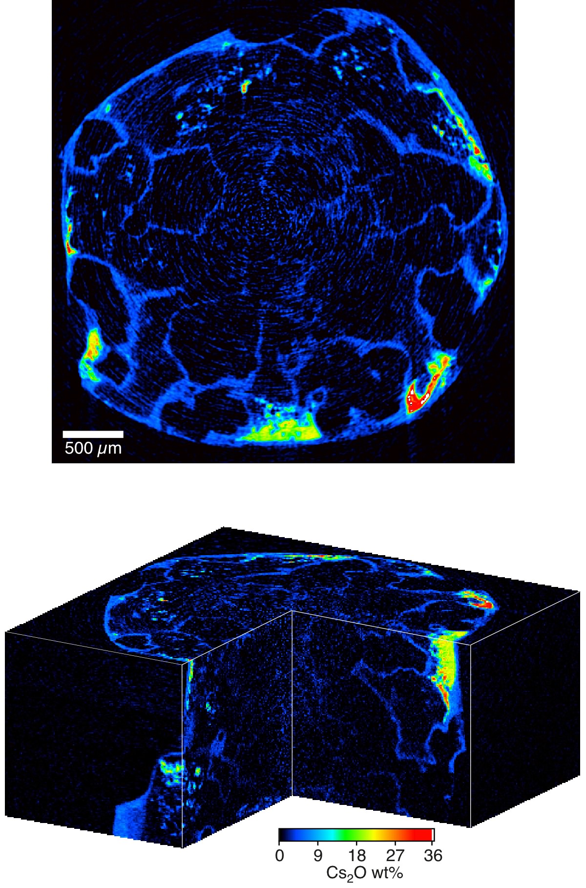
Two-dimensional and three-dimensional maps of Cs2O concentration in the Cs-doped partially molten granite sample
[ S. Ikeda, T. Nakano, A. Tsuchuyama, K. Uesugi, Y. Suzuki, K. Nakamura, Y. Nakashima and H. Yoshida, American Mineralogist 89, 1304-1313 (2004), Fig. 3(b), 3(f),
©2004 Mineralogical Society of America ]
画像ファイルの出典
原著論文/解説記事
誌名
S. Ikeda et al., Am. Mineral. 89, 1304 (2004)
図番号
測定手法
画像ファイルの出典
図なし
測定準備に必要なおおよその時間
1 シフト
測定装置
| 装置名 | 目的 | 性能 |
|---|---|---|
| X-ray CT system | obtain internal structure of material | spatial resolution of 10um |
参考文献
| 文献名 |
|---|
| S. Ikeda et al., Am. Mineral. 89, 1304 (2004) |
関連する手法
アンケート
SPring-8だからできた測定。他の施設では不可能もしくは難しい
本ビームラインの主力装置を使っている
測定の難易度
中程度
データ解析の難易度
中程度
図に示した全てのデータを取るのにかかったシフト数
2~3シフト