X-ray microbeam diffraction (X-ray micro-diffraction)

問い合わせ番号
SOL-0000000927
ビームライン
BL47XU(マイクロCT)
学術利用キーワード
| A. 試料 | 有機材料, 原子・分子・ラジカル, 生物・医学, 計測法、装置に関する研究 |
|---|---|
| B. 試料詳細 | 高分子有機材料, 結晶, 溶液, 液晶, 脂質, 膜, 分子 (中性), 生体高分子、結晶, 蛋白質, 医薬品 |
| C. 手法 | X線回折 |
| D. 手法の詳細 | 単結晶構造解析, 粉末結晶構造解析, 小角散乱 |
| E. 付加的測定条件 | マイクロビーム(<1μm), 二次元画像計測, 応力付加、変形 |
| F. エネルギー領域 | X線(4~40 keV) |
| G. 目的・欲しい情報 | 分子構造, 構造解析, 結晶構造, 欠陥、転位、歪み, 構造変化 |
産業利用キーワード
| 階層1 | 環境, 製薬 |
|---|---|
| 階層2 | ドラッグデザイン, 製剤, 繊維 |
| 階層3 | タンパク質, 薬物, 錠剤 |
| 階層4 | 結晶構造, 配向, 結晶化度, 結晶多形, 内部構造 |
| 階層5 | 回折, X線散乱, 小角散乱, イメージング |
分類
A60.20 環境物質, A80.32 有機材料, A80.40 環境材料, A80.50 製薬, M10.10 単結晶回折, M10.20 粉末結晶回折, M10.30 表面・界面構造回折, M20.10 小角散乱
利用事例本文
X-ray microbeam diffraction (x-ray micro-diffraction) is an accurate technique to study crystal structures of very minute area with sub-micrometer level. Using this technique, one can measure molecular structures in a selected area of sample. Comparing with conventional x-ray diffraction, this technique has other following advantages;
- selectable exposure point
- available for very small crystal
- low background level
- reduce x-ray damage by moving the probe during exposure.
The figure shows the series of x-ray micro-diffraction diagrams of a fiber of high-strength biodegradable polyesters (poly[R)-3-hydroxybutyrate], 50 micron diameter) scanned perpendicular to the fiber axis with a step of 2 microns. Red arrows indicate a new reflection derived from different structure from other parts. These data reveal the fact that this fiber has core-and-sheath structure, with only helix conformation in sheath region and with both planar-zigzag conformation and helix conformation in core region.
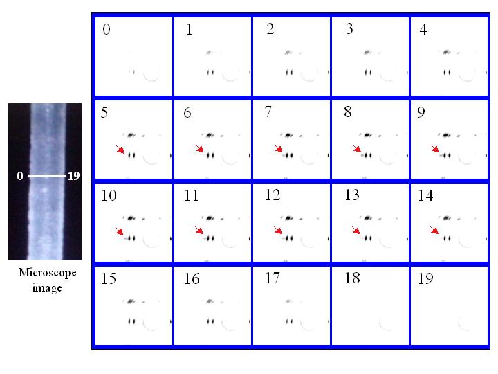
Figure: X-ray micro-diffraction diagrams of fiber of biodegradable polyesters (poly[R)-3-hydroxybutyrate], 50 micron diameter) recorded from the line area in microscope image (left picture).
[ T. Iwata, Y. Aoyagi, M. Fujita, H. Yamane, Y. Doi, Y. Suzuki, A. Takeuchi and K. Ursugi, Macromolecular Rapid Communications 25, 1100-1104 (2004), Fig. 3,
©2004 Wiley VCH ]
画像ファイルの出典
私信等、その他
詳細
理化学研究所 土肥高分子化学研究室の岩田忠久博士
測定手法
Micro-diffraction experiment is performed by using a micro-focused beam generated with a Fresnel zone plate optic. In this solution, diffraction pattern from a small portion (~sub-micrometer) of a sample can be obtained.
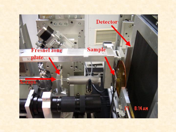
Experimental setup for x-ray micro-diffraction.
画像ファイルの出典
私信等、その他
詳細
理化学研究所 岩田忠久博士
測定準備に必要なおおよその時間
1 日
測定装置
| 装置名 | 目的 | 性能 |
|---|---|---|
| Fresnel zone plate | x-ray micro-focusing | 0.25 micron theoretical resolution |
| Beam monitor 2 | optical alignment | 4.3 micron pixel size |
| Image intensifier | detector | 4 inch field of view |
参考文献
| 文献名 |
|---|
| T. Iwata et. al., Macromol. Rapid Commun. 25, 1100-1104, 2004 |
関連する手法
アンケート
SPring-8だからできた測定。他の施設では不可能もしくは難しい
測定の難易度
中程度
データ解析の難易度
中程度
図に示した全てのデータを取るのにかかったシフト数
2~3シフト